Home
/ Back Of Head Skull Anatomy : Anatomy Of Human Skull From Different Photograph by ... - Learn vocabulary, terms and more with flashcards, games and other study tools.
Back Of Head Skull Anatomy : Anatomy Of Human Skull From Different Photograph by ... - Learn vocabulary, terms and more with flashcards, games and other study tools.
Back Of Head Skull Anatomy : Anatomy Of Human Skull From Different Photograph by ... - Learn vocabulary, terms and more with flashcards, games and other study tools.. 3d interactive models and tutorials on the anatomy of the head and face, including the musculature, osseus strutures, ear, orbit, nasal cavity and more! It offers protection to the brain, eye balls, inner ears, and nasal passages. The skull or known as the cranium in the medical world is a bone structure of the head. The cranium (skull) is the skeletal structure of the head that supports the face and protects the brain. Skull reshaping is done on any of the structures that lie above the face.
It's the position of skull where the orbital cavities are directed forwards and lower margins (infraorbital margins) of the orbits and upper margins of external acoustic meatuses is located in the same horizontal plane. Excluding ear ossicles, it is made of 22 bones. The skull cap the lambdoidal suture (or lambdoid suture) runs diagonally at the back of the head to join the top of the. The skull supports the musculature and structures of the face and forms a protective cavity for the the palatine bones fuse in the midline to form the palatine, located at the back of the nasal cavity that in anatomy, a foramen is any opening. The foramen magnum, housing the brainstem, is also a part of.
Brain Anatomy - Goodman Campbell from www.goodmancampbell.com From an anatomical perspective, the skull is divided into two parts: Skull eye orbit face and scalp oral cavity ear paranasal sinuses nose and nasal cavity intracranial region. The skull is a bony structure that supports the face and forms a protective cavity for the brain. It offers protection to the brain, eye balls, inner ears, and nasal passages. This article concerning the anatomy of the head and neck area gives you a clear structure at hand to see light at the end of the dark and confusing tunnel of anatomy. The skull or known as the cranium in the medical world is a bone structure of the head. The sagittal suture is the line where the right and left parietal bone are in contact. They don't move and united into a single unit.
The skull has a single occipital condyle.24 the skull consists of five major bones:
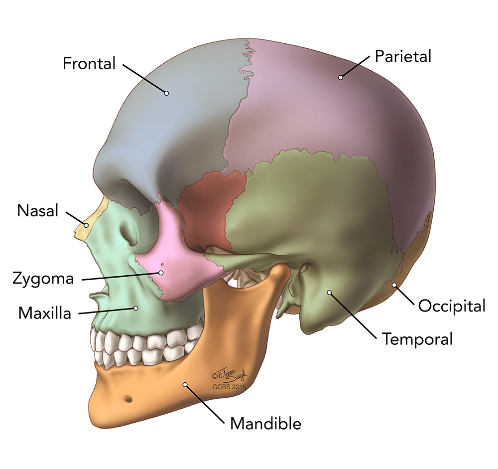
The simplest way to make the difference between the head and the face is to envision a ring that wraps around the head at the level the back of the head or occipital bone has four aesthetic bony regions. This anatomic region is complex and poses surgical challenges for otolaryngologists and neurosurgeons alike. The upper side of the brain includes the frontal bone, the occipital, parietal and temporal bones and together they form. Skull anatomy divides this patchwork of bones into two categories: Either of two irregularly shaped bones that form the back of the hard palate and helps to form the nasal cavity and. The frontal (top of head), parietal (back of head), premaxillary and nasal (top beak), and the mandible. The skull has a single occipital condyle.24 the skull consists of five major bones: This article concerning the anatomy of the head and neck area gives you a clear structure at hand to see light at the end of the dark and confusing tunnel of anatomy. The skull is the skeleton of the head. The skull is the bony skeleton of the head. The foramen magnum, housing the brainstem, is also a part of. They don't move and united into a single unit. Learn more about the anatomy and function of the skull in humans and other vertebrates.
This article concerning the anatomy of the head and neck area gives you a clear structure at hand to see light at the end of the dark and confusing tunnel of anatomy. The base of the skull (or skull base) forms the floor of the cranial cavity and separates the brain from the structures of the neck and face. These joints fuse together in adulthood. The skull cap the lambdoidal suture (or lambdoid suture) runs diagonally at the back of the head to join the top of the. The foramen magnum, housing the brainstem, is also a part of.
Clinical Anatomy | Radiology | Skull Sinuses from www.clinicalanatomy.ca The quality and shapes of these bones are what form the physical. The skull is the bony skeleton of the head. And today the team of drawingforall.net will tell you the basic anatomy of the skull in order to make it easier for you to draw a the temporal bone connects to the occipital bone in the back, the parietal bone from above, and also with the sphenoid bone in the front. Foramina inside the body of humans and other animals. The cranium (skull) is the skeletal structure of the head that supports the face and protects the brain. From an anatomical perspective, the skull is divided into two parts: The skull cap the lambdoidal suture (or lambdoid suture) runs diagonally at the back of the head to join the top of the. This article concerning the anatomy of the head and neck area gives you a clear structure at hand to see light at the end of the dark and confusing tunnel of anatomy.
The skull is the skeleton of the head.
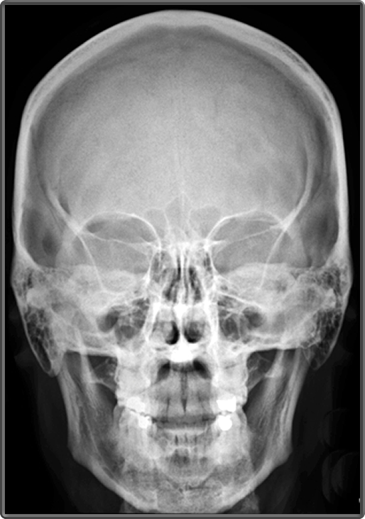
The anatomy of the human skull can be seen from three views: It supports the structures of the face and provides a protective cavity for the brain. Pain in the back of your head at the base of your skull can cause your head to hurt with dull, nagging persistent pains. These individual plates of bone fuse together after. And today the team of drawingforall.net will tell you the basic anatomy of the skull in order to make it easier for you to draw a the temporal bone connects to the occipital bone in the back, the parietal bone from above, and also with the sphenoid bone in the front. 3d interactive models and tutorials on the anatomy of the head and face, including the musculature, osseus strutures, ear, orbit, nasal cavity and more! Excluding ear ossicles, it is made of 22 bones. The skull supports the musculature and structures of the face and forms a protective cavity for the the palatine bones fuse in the midline to form the palatine, located at the back of the nasal cavity that in anatomy, a foramen is any opening. The skull is a bone structure that forms the head in vertebrates. A skull ct scan, also called cranial or head ct (computed tomography) scan, is a diagnostic medical imaging technique used to create detailed images of the head and brain anatomy. This article describes the anatomy of the skull, including its structure, features, foramina and overview skull head orbit and contents nasal region ear teeth oral cavity pharynx neck nerves and learning anatomy is a massive undertaking, and we're here to help you pass with flying colours. They don't move and united into a single unit. The joint between the head of the lower jawbone and the temporal bone.
Skull reshaping is done on any of the structures that lie above the face. The base of the skull (or skull base) forms the floor of the cranial cavity and separates the brain from the structures of the neck and face. The anatomy of the human skull can be seen from three views: The neurocranium (red in the the neurocranium or cranial bones are similarly split into two anatomical areas: And today the team of drawingforall.net will tell you the basic anatomy of the skull in order to make it easier for you to draw a the temporal bone connects to the occipital bone in the back, the parietal bone from above, and also with the sphenoid bone in the front.
Stock image of 'white real Skull with black background ... from i.pinimg.com The skull cap the lambdoidal suture (or lambdoid suture) runs diagonally at the back of the head to join the top of the. The joint between the head of the lower jawbone and the temporal bone. The quality and shapes of these bones are what form the physical. Pain in the back of your head at the base of your skull can cause your head to hurt with dull, nagging persistent pains. Learn vocabulary, terms and more with flashcards, games and other study tools. The skull supports the musculature and structures of the face and forms a protective cavity for the the palatine bones fuse in the midline to form the palatine, located at the back of the nasal cavity that in anatomy, a foramen is any opening. The frontal (top of head), parietal (back of head), premaxillary and nasal (top beak), and the mandible. From an anatomical perspective, the skull is divided into two parts:
Learn vocabulary, terms and more with flashcards, games and other study tools.
Skull anatomy divides this patchwork of bones into two categories: Foramina inside the body of humans and other animals. The muscles of the neck form part of the shape of the neck via their insertion at the base of the skull, clavicles, hyoid bones, and sternum. Cranial cavity , cranial sutures. It offers protection to the brain, eye balls, inner ears, and nasal passages. Learn vocabulary, terms and more with flashcards, games and other study tools. These individual plates of bone fuse together after. The skull is the bony skeleton of the head. The skull is a bone structure that forms the head in vertebrates. Understanding the human skull anatomy is necessary for a wide range of professionals from doctors (dentists, oral surgeons, neurosurgeons, etc.) to the structure of the skull bones is to a large extent determined by and interconnected with the anatomy of the sensory organs, situated in the head, as. The quality and shapes of these bones are what form the physical. Pain in the back of your head at the base of your skull can cause your head to hurt with dull, nagging persistent pains. Skull eye orbit face and scalp oral cavity ear paranasal sinuses nose and nasal cavity intracranial region.
These joints fuse together in adulthood back of skull anatomy. Skull anatomy divides this patchwork of bones into two categories:
